Scientific Programme
Physiology & Nutrition
IS-PN03 - From Sea to Space - Human Physiology and Performance in the Extreme Environments Underwater, High-Altitude, and Weightlessness.
Date: 02.07.2025, Time: 13:15 - 14:30, Session Room: Castello 2
Description
Operating under extreme environmental conditions in recreational, professional, or sports contexts poses significant implications for human physiology, health, and performance. This symposium discusses environmental stressors of underwater, high-altitude, and spaceflight, highlighting acute and long-term adaptations, performance limitations, and coping strategies. Shared risks across these environments include hypoxia, hypercapnia, and decompression due to altered ambient pressure and available life-support solutions, fluid shifts and their effects on gas exchange and work of breathing, and the risk of altered pressures for disease in mountaineers, divers, and astronauts. This symposium highlights risks and potential coping strategies that can be applied before or during exposure to maintain health and performance. Emphasis will be placed on interdisciplinary approaches to enhance human resilience and functionality across these extreme environments, innovative countermeasures, and current limitations.
Chair(s)

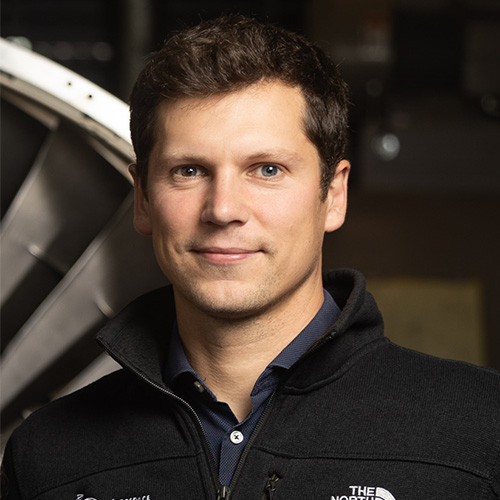
Fabian Möller
Massachusetts Institute of Technology, Aeronautics and Astronautics
United States

Speaker A
Gerardo Bosco
University of Padova, Biomedical Sciences
Italy
Read CVECSS Rimini 2025: IS-PN03
Arterial Partial Pressure of Oxygen Variations in Breath-Hold and SCUBA Diving: State-of-the-Art
According to Boyle’s law and current diving physiology, divers’ arterial blood gas (ABG) levels vary proportionally to environmental pressure underwater. As experimentally demonstrated in hyperbaric chambers, the arterial partial pressures of O2 (PaO2) in SCUBA divers increase during the descent and progressively normalize when resurfacing. In breath-hold divers (BHDs) instead, PaO2 rises during the descent - as lungs compress - but progressively falls when resurfacing due to metabolic consumption and redistribution in re-expanding lungs. This work summarizes the PaO2 from ABGs obtained in the last 4 experimental campaigns on BHDs and SCUBA divers in the real underwater environment. All the experiments were approved by the institution’s ethics committee and included healthy, expert SCUBA divers or BHDs. After inclusion, an arterial cannula was inserted into the radial artery of the non-dominant hand. The ABGs were drawn in different conditions: resting at the surface after cannulation; immediately before diving, after controlled ventilation, in water, face submerged; at depth (-15 mfw or -42 mfw), with BHDs using a sled or fins, and SCUBA divers before and after pedaling on a bike; at surface (after -15 mfw or -42 mfw dives). A scatter plot was used to describe PaO2 distribution in different protocols. Lung ecography have been performed at depth. 34 subjects were included in the study (8 for SCUBA diving), and 150 samples were obtained. Different protocols were applied during these experiments, explaining the variability among the PaO2 results. When at rest, out of the water, all the subjects showed to mirror physiological data. After the pre-dive controlled ventilation, a slight increase can be noted, probably due to water compressing the chest and the lungs of BHDs. Five BHDs out of 14 at -15mfw and 6 out of 20 at -42 mfw did not develop the predicted hyperoxemia at depth, potentially due to lung atelectasis or interstitial edema as reported by lung ecography. At the surface, PaO2 values diminished, with the lowest measured on BHDs using fins to dive. SCUBA divers developed the predicted hyperoxemia at depth without variations after pedaling on the submerged bikes and returned to normal values at the surface. PaO2 in SCUBA divers seems to follow diving physiology predictions; instead, BHDs show higher variability, probably due to environmental stress exerted on the lungs, resulting in lung edema or atelectasis, which affects gas exchange.

Speaker B
Hannah Caldwell
University of Copenhagen, Nutrition, Exercise and Sports (NEXS)
Denmark
Read CVECSS Rimini 2025: IS-PN03
Does maximal exercise at high-altitude exacerbate the brain’s inflammatory response?
The interplay between hypoxia and exercise on the immune response is complex [1,2]. For example, the anti-inflammatory and immunosuppressive effects of strenuous exercise [3] may play a role in immune system sensitization and prevalence of infection at high-altitude [4,5]. Further, pro-inflammatory stress at high-altitude is implicated in the etiology of high-altitude illnesses [6,7]; however, it is controversial whether/how exercise alters pro-inflammatory mediators and therefore the implications of exercise on high-altitude illnesses. How acclimatization to high-altitude affects immune sensitization/suppression and hypersensitivity of inflammatory responses in the brain has not been investigated, particularly following maximal exercise. This talk will discuss the brain’s inflammatory response to both ‘practical’ cycling exercise at a low intensity and in response to acute exhaustive cycling at sea level and following 6-8 days at 3,800 m. Healthy adults (n=12, 6/6 females/males) received radial arterial and internal jugular venous catheterizations to assess trans-cerebral exchange of inflammatory cytokines. Data were obtained prior to and following 60 minutes of steady-state semi-recumbent cycling at an altitude-specific intensity to elicit maximal fat oxidation, and at 5 minutes post- an incremental maximal exercise test. These results indicate that high-altitude per se reduces systemic pro-inflammatory cytokines at rest. Further, 6-8 days of acclimatization at 3,800 m does not provoke immune suppression or mediate elevations in systemic pro-inflammation in response to maximal exercise. Although acute maximal exercise per se does not provoke inflammatory stress in the brain, it appears that the additive duration/intensity effect of prolonged low-intensity exercise followed by acute maximal exercise – irrespective of altitude – provokes a shift toward trans-cerebral net release of pro-inflammatory cytokines. Exposure to high-altitude does not exacerbate pro-inflammatory stress in the brain in response to maximal exercise. Although not directly tested, the current exercise intervention did not initiate any symptoms of altitude illness or headache. As pro-inflammatory stress is implicated in the pathophysiology of altitude illness [6,7], these data support that, in the context of the current experimental study following acclimatization to 3,800 m, this duration and intensity of exercise is not a trigger for altitude illness via pro-inflammatory stress. These data provide insights into the tolerability of acute maximal exercise at high-altitude. [1] Vats et al. (2006). DOI: 10.1016/j.cmet.2006.05.011. [2] Weinberg et al. (2015). DOI: 10.1016/j.immuni.2015.02.002. [3] Shaw et al. (2018). DOI: 10.1016/j.cyto.2017.10.001. [4] Facco et al. (2005). DOI: 10.1249/01.mss.0000162688.54089.ce. [5] Feuerecker et al. (2019). DOI: 10.1111/all.13545. [6] Pham et al. (2021). DOI: 10.3389/fphys.2021.676782. [7] Turner et al. (2021). DOI: 10.1152/japplphysiol.00861

Speaker C
Fabian Möller
Massachusetts Institute of Technology, Aeronautics and Astronautics
United States
Read CVECSS Rimini 2025: IS-PN03
Spaceflight Physiology: The Role of Countermeasures for Health and Performance in Weightlessness
Prerequisites for human performance during spaceflight are survival in the extreme space environment and solutions to maintain health in weightlessness. Habitat and life-support solutions keep astronauts alive against the imminent threats of vacuum, temperature, and carbon dioxide accumulation, and countermeasures are implemented to maintain health and attenuate cardiovascular and musculoskeletal deconditioning. Specific risks associated with weightlessness, such as hypoxia, hypercapnia, and changes in pressure, are similar to those encountered in underwater and high-altitude environments. This talk discusses the physiological adaptations in weightlessness and the role of countermeasures and gravitational stress in maintaining health and performance during spaceflight. Current plans of national space agencies and commercial providers will increase the number of people in space, and tasks like extra-vehicular activities, the return to a gravitational field, and future planetary explorations demand intact high performance to ensure safety and mission success. Current countermeasures include engineering, medical, and exercise interventions, but astronauts continue to return with impaired vision and losses in bone mineral density, muscle mass, and strength [1–3]. Determining the necessary amount of gravity to maintain astronaut health remains a critical challenge. While 1G on Earth appears optimal, it is unclear how much gravitational stress is needed in space to achieve similar health outcomes. Weightlessness induces a cranial fluid shift with central hypervolemia, increased intracranial and intraocular pressures, and reduced venous return [4], suspected to cause the observed ocular morphology and vision changes during and following long-duration spaceflight [5,6]. Furthermore, the lack of gravitational stress deconditions the cardiovascular and musculoskeletal systems, reducing aerobic capacity and increasing the risk of bone fracture during planetary landings and orthostatic intolerance after returning to a gravitational field. While the International Space Station offers various countermeasures, medical support, and evacuation strategies, future long-duration spaceflight will amplify current risks while posing severe volume and weight limitations, demanding novel and comprehensive countermeasure solutions. [1] Comfort et al. (2021). DOI 10.1007/s40279-021-01496-9 [2] Ong et al. (2023). DOI: 10.1038/s41433-023-02522-y. [3] Scott et al. (2023). DOI: 10.1038/s41526-023-00256-5. [4] Roberts and Petersen (2019). DOI: 10.1001/jamaneurol.2018.4891. [5] Laurie et al. (2020). DOI: 10.1001/jamaophthalmol.2019.5261. [6] Macias et al. (2021). DOI: 10.1001/jamaophthalmol.2021.0931.
